
The mysterious dance of the cricket embryos
New York Times

- +
- -
In June, 100 fruit fly scientists gathered
on the Greek island of Crete for their biennial meeting. Among them was
Cassandra Extavour, a Canadian geneticist at Harvard University. Her lab works
with fruit flies to study evolution and development — “evo devo.” Most often,
such scientists choose as their “model organism” the species Drosophila
melanogaster — a winged workhorse that has served as an insect collaborator on
at least a few Nobel Prizes in physiology and medicine.اضافة اعلان
But Extavour is also known for cultivating alternative species as model organisms. She is especially keen on the cricket, particularly Gryllus bimaculatus, the two-spotted field cricket, even though it does not yet enjoy anything near the fruit fly’s following. (Some 250 principal investigators had applied to attend the meeting in Crete.)
Extavour’s research aims at the fundamentals: How do embryos work? And what might that reveal about how the first animal came to be? Every animal embryo follows a similar journey: One cell becomes many, then they arrange themselves in a layer at the egg’s surface, providing an early blueprint for all adult body parts. But how do embryo cells — cells that have the same genome but are not all doing the same thing with that information — know where to go and what to do?
“That’s the mystery for me,” Extavour said. “That’s always where I want to go.”
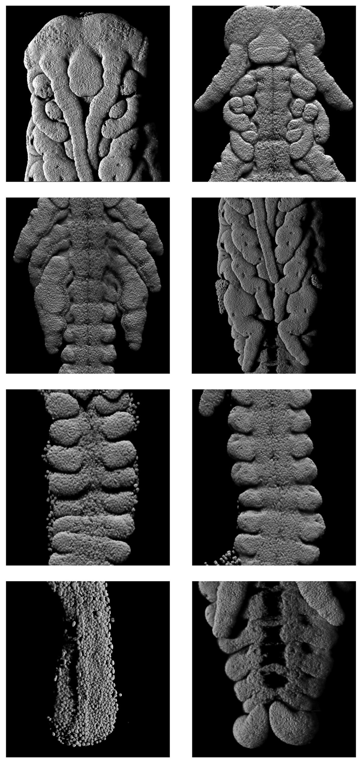
Seth Donoughe, a biologist and data scientist at the University of Chicago and an alumnus of Extavour’s lab, described embryology as the study of how a developing animal makes “the right parts at the right place at the right time.” In some new research featuring wondrous video of the cricket embryo — showing certain “right parts” (the cell nuclei) moving in 3D — Extavour, Donoughe, and their colleagues found that good old-fashioned geometry plays a starring role.
Humans, frogs, and many other widely studied animals start as a single cell that immediately divides again and again into separate cells. In crickets and most other insects, initially just the cell nucleus divides, forming many nuclei that travel throughout the shared cytoplasm and only later form cellular membranes of their own.
In 2019, Stefano Di Talia, a quantitative developmental biologist at Duke University, studied the movement of the nuclei in the fruit fly and showed that they are carried along by pulsing flows in the cytoplasm — a bit like leaves traveling on the eddies of a slow-moving stream.
But some other mechanism was at work in the cricket embryo. The researchers spent hours watching and analyzing the microscopic dance of nuclei: glowing nubs dividing and moving in a puzzling pattern, not altogether orderly, not quite random, at varying directions and speeds, neighboring nuclei more in sync than those farther away. The performance belied a choreography beyond mere physics or chemistry.
“The geometries that the nuclei come to assume are the result of their ability to sense and respond to the density of other nuclei near to them,” Extavour said. Di Talia was not involved in the new study but found it moving. “It’s a beautiful study of a beautiful system of great biological relevance,” he said.
Journey of the nuclei
The cricket researchers at first took a classic approach: Look closely and pay attention. “We just watched it,” Extavour said.
They shot videos using a laser-light sheet microscope: Snapshots captured the dance of the nuclei every 90 seconds during the embryo’s initial eight hours of development, in which time 500 or so nuclei had amassed in the cytoplasm. (Crickets hatch after about two weeks.)
Initially the biologists watched the videos on loop, projected onto a conference-room screen — the cricket-equivalent of IMAX, considering that the embryos are about one-third the size of a grain of (long-grain) rice. They tried to detect patterns, but the data sets were overwhelming. They needed more quantitative savvy.
Donoughe contacted Christopher Rycroft, an applied mathematician now at the University of Wisconsin-Madison, and showed him the dancing nuclei. “Wow!” Rycroft said. He had never seen anything like it, but he recognized the potential for a data-powered collaboration; he and Jordan Hoffmann, then a doctoral student in Rycroft’s lab, joined the study.
Over numerous screenings, the math-bio team contemplated many questions: How many nuclei were there? When did they start to divide? What directions were they going in? Where did they end up? Why were some zipping around and others crawling?
Rycroft often works at the crossroads of the life and physical sciences. (Last year, he published on the physics of paper crumpling.) “Math and physics have had a lot of success in deriving general rules that apply broadly, and this approach may also help in biology,” he said; Extavour has said the same.
The team spent a lot of time swirling ideas around at a white board, often drawing pictures. The problem reminded Rycroft of a Voronoi diagram, a geometric construction that divides a space into nonoverlapping subregions — polygons, or Voronoi cells, that each emanate from a seed point. It’s a versatile concept that applies to things as varied as galaxy clusters, wireless networks and the growth pattern of forest canopies. (The tree trunks are the seed points and the crowns are the Voronoi cells, snuggling closely but not encroaching on one another, a phenomenon known as crown shyness.)
In the cricket context, the researchers computed the Voronoi cell surrounding each nucleus and observed that the cell’s shape helped predict the direction the nucleus would move next. Basically, Donoughe said, “Nuclei tended to move into nearby open space.”
Geometry, he noted, offers an abstracted way of thinking about cellular mechanics. “For most of the history of cell biology, we couldn’t directly measure or observe the mechanical forces,” he said, even though it was clear that “motors and squishes and pushes” were at play. But researchers could observe higher-order geometric patterns produced by these cellular dynamics. “So, thinking about the spacing of cells, the sizes of cells, the shapes of cells — we know they come from mechanical constraints at very fine scales,” Donoughe said.
To extract this sort of geometric information from the cricket videos, Donoughe and Hoffmann tracked the nuclei step-by-step, measuring location, speed, and direction.
“This is not a trivial process, and it ends up involving a lot of forms of computer vision and machine-learning,” said Hoffmann, an applied mathematician now at DeepMind in London.
They also verified the software’s results manually, clicking through 100,000 positions, linking the nuclei’s lineages through space and time. Hoffmann found it tedious; Donoughe thought of it as playing a video game, “zooming in high-speed through the tiny universe inside a single embryo, stitching together the threads of each nucleus’s journey.”
Next they developed a computational model that tested and compared hypotheses that might explain the nuclei’s motions and positioning. All in all, they ruled out the cytoplasmic flows that Di Talia saw in the fruit fly. They disproved random motion and the notion that nuclei physically pushed each other apart.
Instead, they arrived at a plausible explanation by building on another known mechanism in fruit fly and roundworm embryos: miniature molecular motors in the cytoplasm that extend clusters of microtubules from each nucleus, not unlike a forest canopy.
The team proposed that a similar type of molecular force drew the cricket nuclei into unoccupied space. “The molecules might well be microtubules, but we don’t know that for sure,” Extavour said in an email. “We will have to do more experiments in the future to find out.”
Read more Odd and Bizarre
Jordan News
But Extavour is also known for cultivating alternative species as model organisms. She is especially keen on the cricket, particularly Gryllus bimaculatus, the two-spotted field cricket, even though it does not yet enjoy anything near the fruit fly’s following. (Some 250 principal investigators had applied to attend the meeting in Crete.)
Extavour’s research aims at the fundamentals: How do embryos work? And what might that reveal about how the first animal came to be? Every animal embryo follows a similar journey: One cell becomes many, then they arrange themselves in a layer at the egg’s surface, providing an early blueprint for all adult body parts. But how do embryo cells — cells that have the same genome but are not all doing the same thing with that information — know where to go and what to do?
“That’s the mystery for me,” Extavour said. “That’s always where I want to go.”
Seth Donoughe, a biologist and data scientist at the University of Chicago and an alumnus of Extavour’s lab, described embryology as the study of how a developing animal makes “the right parts at the right place at the right time.” In some new research featuring wondrous video of the cricket embryo — showing certain “right parts” (the cell nuclei) moving in 3D — Extavour, Donoughe, and their colleagues found that good old-fashioned geometry plays a starring role.
Humans, frogs, and many other widely studied animals start as a single cell that immediately divides again and again into separate cells. In crickets and most other insects, initially just the cell nucleus divides, forming many nuclei that travel throughout the shared cytoplasm and only later form cellular membranes of their own.
In 2019, Stefano Di Talia, a quantitative developmental biologist at Duke University, studied the movement of the nuclei in the fruit fly and showed that they are carried along by pulsing flows in the cytoplasm — a bit like leaves traveling on the eddies of a slow-moving stream.
But some other mechanism was at work in the cricket embryo. The researchers spent hours watching and analyzing the microscopic dance of nuclei: glowing nubs dividing and moving in a puzzling pattern, not altogether orderly, not quite random, at varying directions and speeds, neighboring nuclei more in sync than those farther away. The performance belied a choreography beyond mere physics or chemistry.
“The geometries that the nuclei come to assume are the result of their ability to sense and respond to the density of other nuclei near to them,” Extavour said. Di Talia was not involved in the new study but found it moving. “It’s a beautiful study of a beautiful system of great biological relevance,” he said.
Journey of the nuclei
The cricket researchers at first took a classic approach: Look closely and pay attention. “We just watched it,” Extavour said.
They shot videos using a laser-light sheet microscope: Snapshots captured the dance of the nuclei every 90 seconds during the embryo’s initial eight hours of development, in which time 500 or so nuclei had amassed in the cytoplasm. (Crickets hatch after about two weeks.)
Initially the biologists watched the videos on loop, projected onto a conference-room screen — the cricket-equivalent of IMAX, considering that the embryos are about one-third the size of a grain of (long-grain) rice. They tried to detect patterns, but the data sets were overwhelming. They needed more quantitative savvy.
Donoughe contacted Christopher Rycroft, an applied mathematician now at the University of Wisconsin-Madison, and showed him the dancing nuclei. “Wow!” Rycroft said. He had never seen anything like it, but he recognized the potential for a data-powered collaboration; he and Jordan Hoffmann, then a doctoral student in Rycroft’s lab, joined the study.
Over numerous screenings, the math-bio team contemplated many questions: How many nuclei were there? When did they start to divide? What directions were they going in? Where did they end up? Why were some zipping around and others crawling?
Rycroft often works at the crossroads of the life and physical sciences. (Last year, he published on the physics of paper crumpling.) “Math and physics have had a lot of success in deriving general rules that apply broadly, and this approach may also help in biology,” he said; Extavour has said the same.
The team spent a lot of time swirling ideas around at a white board, often drawing pictures. The problem reminded Rycroft of a Voronoi diagram, a geometric construction that divides a space into nonoverlapping subregions — polygons, or Voronoi cells, that each emanate from a seed point. It’s a versatile concept that applies to things as varied as galaxy clusters, wireless networks and the growth pattern of forest canopies. (The tree trunks are the seed points and the crowns are the Voronoi cells, snuggling closely but not encroaching on one another, a phenomenon known as crown shyness.)
In the cricket context, the researchers computed the Voronoi cell surrounding each nucleus and observed that the cell’s shape helped predict the direction the nucleus would move next. Basically, Donoughe said, “Nuclei tended to move into nearby open space.”
Geometry, he noted, offers an abstracted way of thinking about cellular mechanics. “For most of the history of cell biology, we couldn’t directly measure or observe the mechanical forces,” he said, even though it was clear that “motors and squishes and pushes” were at play. But researchers could observe higher-order geometric patterns produced by these cellular dynamics. “So, thinking about the spacing of cells, the sizes of cells, the shapes of cells — we know they come from mechanical constraints at very fine scales,” Donoughe said.
To extract this sort of geometric information from the cricket videos, Donoughe and Hoffmann tracked the nuclei step-by-step, measuring location, speed, and direction.
“This is not a trivial process, and it ends up involving a lot of forms of computer vision and machine-learning,” said Hoffmann, an applied mathematician now at DeepMind in London.
They also verified the software’s results manually, clicking through 100,000 positions, linking the nuclei’s lineages through space and time. Hoffmann found it tedious; Donoughe thought of it as playing a video game, “zooming in high-speed through the tiny universe inside a single embryo, stitching together the threads of each nucleus’s journey.”
Next they developed a computational model that tested and compared hypotheses that might explain the nuclei’s motions and positioning. All in all, they ruled out the cytoplasmic flows that Di Talia saw in the fruit fly. They disproved random motion and the notion that nuclei physically pushed each other apart.
Instead, they arrived at a plausible explanation by building on another known mechanism in fruit fly and roundworm embryos: miniature molecular motors in the cytoplasm that extend clusters of microtubules from each nucleus, not unlike a forest canopy.
The team proposed that a similar type of molecular force drew the cricket nuclei into unoccupied space. “The molecules might well be microtubules, but we don’t know that for sure,” Extavour said in an email. “We will have to do more experiments in the future to find out.”
Read more Odd and Bizarre
Jordan News